
I am studying Westheide und Rieger's "Spezielle Zoologie" for several days now and I am amazingly surprised of the variability and number of taxa having parasitic lifestyles. Here’s about crustacean especially. But I am quite certain that other taxa will make me post as well.
Prof. H.K. Schminke from the University of Oldenburg, Germany wrote the chapter about Crustaceans. He keeps using a highly unresolved class called Maxillopoda to put in a couple of subtaxa which can so far not be linked to other Crustacean groups but are alltogether most likely no monophyletic group. Crustaceans most of them are. I am not joking. Included are coequally 9 subtaxa, namely the Mystacocarida, Copepoda, Branchiura (Fish Lice), Pentastomida (Tongue Worms), Ostracoda (Seed Shrimps), Tantulocarida, Facetotecta, Ascothoracida and Cirripedia (Barnacles and relatives). Other sources like the "Tree of Life web project" and some authors cited therein sum up Cirripedia, Facetotecta and Ascothoracida to a group called Thecostraca. But this doesn’t matter here. Fact is that some of these taxa have been mysteries until recently, at least for systematic zoologists.
The Facetotecta for example have until this year only been known because of their derived larvae, called y-nauplius (5 stages) and y-cypris (1 stage). These have been found worldwide and do not seem to be rare. Even though adult stages were never found so far, some larvae have been described as species and all the taxonomy and further systematic research on this group are based solely on the larvae. All described species belong to the genus Hansenocaris. The y-cypris has an undivided carapace and a “lattice organ”. The latter is most likely a chemo-sensory organ and links Facetotects to the barnacles and relatives. Its only extremities are the 1. antennae of which the 2nd segment may bear retractable claws. The labrum is long and also bears up to five hook-like claws covering the ventral cephalon. 6 pairs of thoracopods are made for swimming and the cypris has both, a single naupliar eye and a single compound eye. These appendages always were used to hypothesize a parasitic adult stage.
By reading this very interesting Blog by Christopher Taylor I was just pointed to a relatively new publication by Glenner et al. 2008 who found this previously unknown adult of a y-larva by induced metamorphosis. Well, it is no surprise that this adult stage of the so called Facetotecta was previously unknown despite the fact the its y-nauplius larva was known since the end of the 19th century. The authors point out that the animal is not capable of living on its own after metamorphosis and conclude it must be a parasite. But still nobody knows were to find the host. Isn’t that amazing? This kind of riddles is the reason for me to study biology and to do marine research, especially on crustaceans. Many mysteries remain unsolved!
More meshugga lifestyles: While barnacle-like cirripeds spend their adult life as sessile organisms on inorganic substrate (Thoracica) or buried in limestone or calcareous shells (Acrothoracica) filtering water, the Rhizocephala are a sistertaxon with a completely different lifestyle and another miracle of parasitism. Their relationship to other crustaceans and especially cirripeds was clarified by discovering their larval stages. But even to identify the adults as crustaceans is not possible on a morphological basis. Around
Female cypris larvae have to find the host – crabs in general. They penetrate soft parts of the Cuticula, usually joints on the ventral side of the pleon. These joints are not calcified and not sklerotized. When molting, the larvae grow into the host’s body. On the outside of the hosts cuticle a sack develops (Externa) as the posterior part and breeding organ of the parasite. Inside the host a root-system like anterior part (Interna) braids the internal organs of the host. Through this Interna the parasite feeds and also infers with the hormone system of the crab. The latter is sterilized and changes its body proportions (e.g. female-like pleons form in males).
Male rhizocephalans are dwarf-like and look different from the females even as larvae. After finding the mate, males swim to the Externa. Here they attach and leave their Cuticula - through the first Antennae which open at predetermined breaking points - as extremely small males. These are called Trichogons or dwarf males. The Trichogons become ameboid and penetrate the externa. By means of peristaltic movements they are moved to some receptacle, molt again and therewith close the entrance to keep away other males. The male is now implanting itself to the female body and starts producing sperm after some days. A female features two of these receptacles and can therefore incorporate two males. They will continue producing offspring together for a whole lifetime.
The male is nothing more but a sperm-producing amoeba living inside its mate. They invest 100% of “their” energy in sperm. The female keeps up to two males feeding them. 50% of her body is dedicated to rearing eggs and the other 50% are for manipulating the crab, which, male or female, form a broad Pleon as typical for females, not to take care of its own offspring but of the offspring of the parasites. Unbelievable!
In some other species the male larvae can inject their Trichogon through the cuticle of the female or even through the cuticle of the host when the female is located underneath. They accomplish that by means of their Antennae which they can use as an injection needle. You can find information about host specificity and an illustrated lifecycle of Sacculina carcini in a paper of Goddard et al. 2005. Well, it is especially about hosts in areas where it is found as an invader but still..
The Pentastomida are another subclass of “Maxillopoda” with not only obligatory parasitic stages but complete parasitic lifecycles. And this group was a riddle, too, as it was controversial for a long time, in which relationship to see them. They do not even have typical larvae to allocate them to crustaceans. And as the colloquial name “tongue worm” indicates their body plan is rather distinct from all other arthropods and even wormlike taxa among the invertebrates have not much in common with tongue worms.
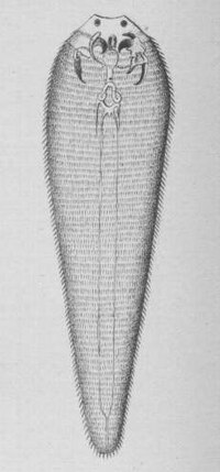
Tongue worms get up to 16cm in length and have a highly degenerated morphology. Adults live as ectoparasites in lungs, paranasal sinuses and trachea of terrestrial carnivore and omnivore vertebrates. Here they suck blood and mucus. 90% breed in reptiles, others in Canidae and Aves. The larvae live in many different terrestrial vertebrates of all groups and some Blattodea and Coleoptera. Linguatula serrata is common in dogs and may accidentally infect humans.
The latin name was caused by a misinterpretation of two pairs of openings next to the mouth opening comprising a retractable claw each. These claws may be homologous to Mandible and first Maxille in Arthropods. Except of additional frontal and apical papillae, which are interpreted as sensory system, there are no other appendages. There is no heart. Haemolymph is pumped by peristaltic movement of the whole body. There is no organ for excretion. The Body is annulated up to 230 times with two pairs of dorsoventral muscles for each annulus. This indicates a real metamerism. These are special developments which possibly are apomorphic characters for pentastomids. But these characters can be found nowhere else in crustaceans!
The Embryo has 4 pairs of appendage buds and a dorsal organ producing mucus. The four anterior buds develop to the frontal and apical papillae and the others make the claws. Anterior to the mouth the larva has a stiletto to penetrate the intestine wall of the host. Once it has found an alternate host or reinfected the original host, it moves through the body feeding and moulting several times. It develops to the adult stage gradually or another second maggot-like type of larvae may be interconnected. Alternate hosts normally are prey for the potential hosts.
Many works tried to find similarities between Pentastomida and any phylum of the animal kingdom or at least tried to eliminate possibilities. For example Karuppaswamy (1976) found β-Chitin in the cuticle of the Pentastomid Raillietiella gowrii using an X-ray technique and therefore delimited Pentastomulida from Arthropoda.
But finally, Lavrov et al. (2004) looked at the complete mtDNA sequence and gene arrangements and found a relation to Maxillopoda and especially Branchiura. This hypothesis remains to be tested. However, fossils from the Upper Cambrian identified as pentastomids (Walossek, D. and K. J. Müller, 1998) indicate an early branch dividing Pentastomida from the other Arthropods.
Whatsoever. Work remains to be done.
References:
- Walossek, D. and K. J. Müller (1998). "Cambrian "Orsten"-type arthropods and the phylogeny of Crustacea” in R. A. Fortey and R. H. Thomas: Arthropod Relationships. London: Chapman & Hall, 139–143.
- Goddard, J.H.R., M.E. Torchin, A.M. Kuris & K.D. Lafferty (2005); Host specificity of Sacculina carcini, a potential biological control agent of the introduced European green crab Carcinus maenas in
- Glenner, H., J.T. Høeg, M.J. Grygier & Y. Fujita (2008); Induced metamorphosis in crustacean y-larvae: Towards a solution to a 100-year-old riddle. BMC Biology
-
- Schminke,H.K. (2007); „Crustacea“ in Westheide, W. & R. Rieger’s „Spezielle Zoologie Teil 1. Einzeller und Wirbellose. Elsevier. Spektrum Akademischer Verlag 555-637.
-
No comments:
Post a Comment